
SPECTROSCOPY SYSTEMS:
Confocal Raman Spectrometer
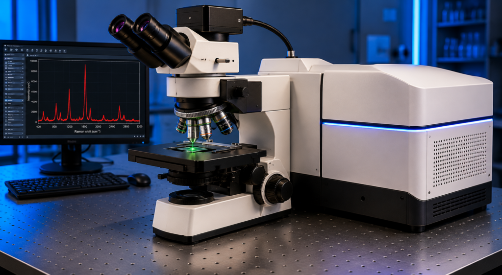
Principle:
Raman spectroscopy is based on the inelastic scattering of monochromatic light (laser) by molecules.
- When a laser interacts with a sample, most light undergoes elastic (Rayleigh) scattering with no energy change.
- A small fraction undergoes inelastic (Raman) scattering, where energy is gained or lost due to molecular vibrations.
- The energy shift between incident and scattered light provides a molecular fingerprint.
- This helps identify chemical bonds, functional groups, and structural information.
Key Components:
- Laser source: Provides monochromatic excitation (e.g., 488 nm, 532 nm, 633 nm, 785 nm)
- Confocal microscope: Enables high spatial resolution and depth profiling
- Spectrometer: Disperses scattered light into Raman spectra
- Detector: CCD/PMT for signal detection
- Motorized stage: For precise sample positioning and mapping
Key Features:
- Confocal capability: Depth-resolved (3D) analysis
- Non-destructive technique
- Minimal sample preparation required
- High chemical specificity (molecular fingerprinting)
- 2D and 3D Raman mapping
- Multi-mode analysis: Raman, photoluminescence, and reflection measurements
- High spatial resolution imaging
Specifications:
-
Spectral Range (Raman shift):
Typically 50 – 4000 cm⁻¹ (standard range)
Extended capability up to ~6000–10000 cm⁻¹ (depending on laser and configuration) -
Laser Options:
Multiple excitation wavelengths available:
405 nm, 488 nm, 532 nm, 633 nm, 785 nm
1 to 5 lasers can be integrated (model dependent) -
Spectral Resolution:
Typical: 0.5 – 2 cm⁻¹
High-resolution mode (Echelle grating): up to ~0.25 cm⁻¹
Lower-end systems: up to ~4 cm⁻¹ -
Gratings:
Interchangeable gratings:
600, 1200, 1800, 2400 lines/mm
Optional Echelle grating for ultra-high resolution -
Detector:
Thermoelectrically cooled CCD detector (standard)
Optional: PMT / photosensor modules for high-speed -
Spectrometer:
High-throughput spectrometer
Optical range: ~190 – 1100 nm or 400 – 1100 nm -
Focal Length:
Typically, 200 – 750 mm (depends on configuration) -
Spatial Resolution (Confocal):
Lateral (XY): ~0.3 – 1 µm
Axial (Z): ~0.7 – 2 µm -
Scanning / Mapping:
2D and 3D Raman mapping capability
Minimum step size: ~100 nm
Typical scan range (microscopy): ~200 × 200 µm (or higher with stage movement) -
Repeatability:
Approximately ±0.006 to ±0.03 nm -
Additional Modes (in advanced systems):
Photoluminescence (PL)
Rayleigh imaging
CARS (Coherent Anti-Stokes Raman Scattering)
Applications:
-
Chemical Identification :
Identifies API and excipients in pharmaceutical products using Raman spectra. -
Drug–Excipient Interaction Studies:
Detects interactions between drug and excipients in tablets or formulations. -
Raman Chemical Imaging:
Creates 2D or 3D chemical maps showing the distribution of different components in a sample. -
Polymorph Identification:
Distinguishes different polymorphic forms of APIs, which is important for drug stability and bioavailability. -
Particle Analysis:
Determines particle composition and distribution in powders or mixtures. -
Quality Control in Pharmaceuticals
Used to verify uniformity of tablets, capsules, and injectables. -
Non-destructive Analysis:
Analyzes samples without damaging them. -
Surface and Microstructure Analysis:
Studies coatings, layers, and microstructures in materials
Ready to Work With Podhikai Scientific?
Take the next step in strengthening your laboratory or research facility with
reliable scientific instruments. Contact us to discuss your requirements or
learn more about our solutions.