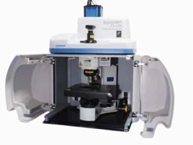
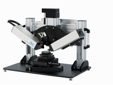
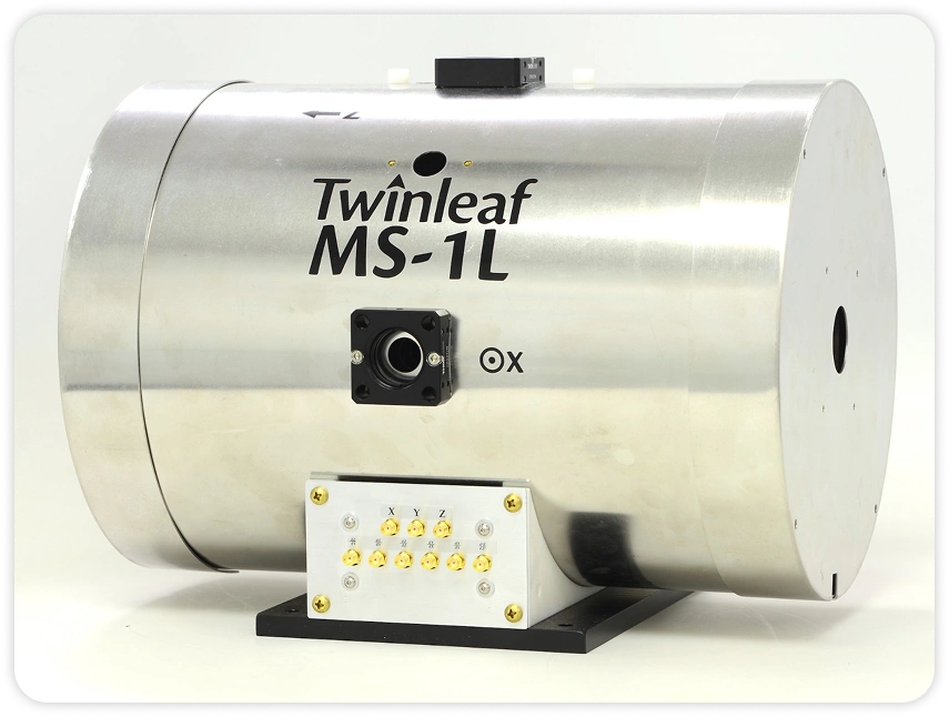
Optical microscope
- Home
- Optical microscope
Types of Techniques
- Atomic Force Microscopy (AFM)
- Field Emission-Scanning Electron Microscopy (FESEM)
- Optical microscope
- Transmission Electron Microscopy (TEM)
- Scanning Acoustic microscopy
- Confocal Micro/Nano Photoluminescence Spectroscopy (PL)
- Confocal micro /nano Raman spectroscopy
- Focused Ion Beam – Scanning Electron Microscopy
- Electron Probe Micro Analysis (EPMA)
- Focused Ion Beam (FIB)
- Infinite Focus Microscopy
- Cathodo lumiscence
Optical microscope

An optical microscope is an instrument that uses visible light and a system of lenses to magnify small objects, allowing them to be observed in greater detail. It is one of the most fundamental and widely used tools in various fields of science, including biology, medicine, materials science, and more.
The basic working principle of an optical microscope involves the use of two types of lenses: the objective lens and the eyepiece lens. The objective lens is the lens closest to the specimen, and it collects light from the object and forms a real, inverted, and magnified image. The eyepiece lens, or ocular lens, acts as a simple magnifying glass that further magnifies the image formed by the objective lens, allowing the user to observe the enlarged image.
Here are the experiments/applications that can be performed using an optical microscope across various industries, presented in a point-wise format:
Aerospace:- Inspection of aircraft components for defects, wear, or damage
- Analysis of materials and coatings used in aerospace applications
- Examination of fuel system components and filters
- Examination of engine components, such as pistons, valves, and cylinders
- Analysis of paint and coatings for quality control
- Inspection of electrical components and wiring
- Analysis of chemical samples for purity and identification of contaminants
- Examination of catalysts and reaction products
- Identification of crystalline structures and polymorphic forms
- Inspection of circuit boards and electronic components for defects or quality control
- Analysis of materials used in consumer electronics products
- Examination of display components and coatings
- Forensic examination of evidence, such as fibers, hair, and gunshot residue
- Analysis of materials used in defense equipment and weapons
- Inspection of chemical and biological samples
- Analysis of materials used in energy production and storage systems
- Examination of fuel samples and contaminants
- Inspection of components in energy generation equipment
- Forensic examination of evidence, such as trace materials, fibers, and biological samples
- Analysis of documents and handwriting for authentication
- Examination of counterfeit products or materials
- Inspection of LED components and materials for quality control
- Analysis of coatings and optical properties of lighting products
- Examination of phosphor materials used in LED manufacturing
- Examination of medical device components and materials for defects or quality control
- Analysis of surface finishes and coatings on medical devices
- Inspection of biological samples and tissues
- Analysis of drug samples for purity and identification of contaminants
- Examination of packaging materials for defects or quality control
- Inspection of raw materials used in pharmaceutical manufacturing
- Inspection of raw materials for defects, impurities, or contaminants
- Analysis of material properties and composition
- Examination of particle size and distribution
- Inspection of semiconductor wafers and components for defects or quality control
- Analysis of materials used in semiconductor manufacturing
- Examination of thin-film coatings and structures
- Examination of electronic components and materials used in telecommunications equipment
- Inspection of optical fibers and connectors
- Analysis of materials used in data storage devices
- Simple and user-friendly operation
- Ability to observe live specimens
- Wide range of magnification (up to 1000x or more)
- Relatively inexpensive compared to other microscopy techniques
- Versatile sample preparation methods
- Samples should be thin and transparent (or semi-transparent) for light transmission
- Biological samples may require staining or mounting on glass slides
- Opaque samples can be observed using reflected light techniques
- Sample size ~ maximum 15 cm2